- Suite 1, Level 1, 117 Camberwell Road, Hawthorn East VIC 3123
- [email protected]


LAM
Lymphangioleiomyomatosis
About LAM
- Home
- About LAM
A rare disease with clear underlying biology
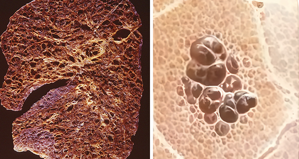
The left-hand section of lung shows extensive cysts (dark red) and destruction of normal lung². In the right-hand picture, cysts are clearly shown within normal lung tissue⁵.
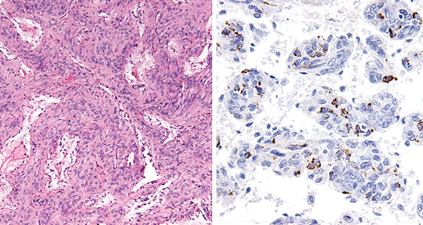
These pictures taken under high power microscopes show abundant proliferation of abnormal smooth muscle–like cells (LAM cells)³˒⁴.
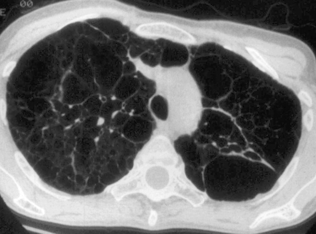
This high-resolution CT scan shows severe cyst infiltration (black pockets) in the lungs².
Over the past two decades, understanding of LAM has changed fundamentally. Rather than being viewed simply as a rare cystic lung disease, LAM is now recognised as a disease of abnormal cell growth. The World Health Organization (WHO) classifies LAM as a low-grade mesenchymal neoplasm within the family of perivascular epithelioid cell tumours (PEComas). This classification reflects the biological behaviour of the abnormal LAM cells and has fundamentally changed the scientific understanding of the disease.
The first biological breakthrough
One of the most important advances in understanding LAM was the discovery that loss of function of the tumour suppressor genes TSC1 or TSC2 drives abnormal LAM cell growth. Under normal circumstances these genes produce two proteins, hamartin and tuberin, which function together as one of the cell’s natural growth-control systems by regulating the mTOR signalling pathway, one of the cell’s master regulators of growth, metabolism, proliferation and survival.
When this control system is lost, the mTOR pathway becomes persistently activated. Abnormal LAM cells survive longer than they should, continue to divide and gradually accumulate throughout the lungs and lymphatic system.
This discovery transformed the treatment of LAM and established the first successful example of targeting one of the disease’s fundamental biological mechanisms rather than simply treating the consequences of lung damage. It also demonstrated that understanding the biology of LAM could lead directly to new therapies and opened the way to identifying additional biological pathways that may contribute to disease progression beyond mTOR.
One existing treatment with limitations
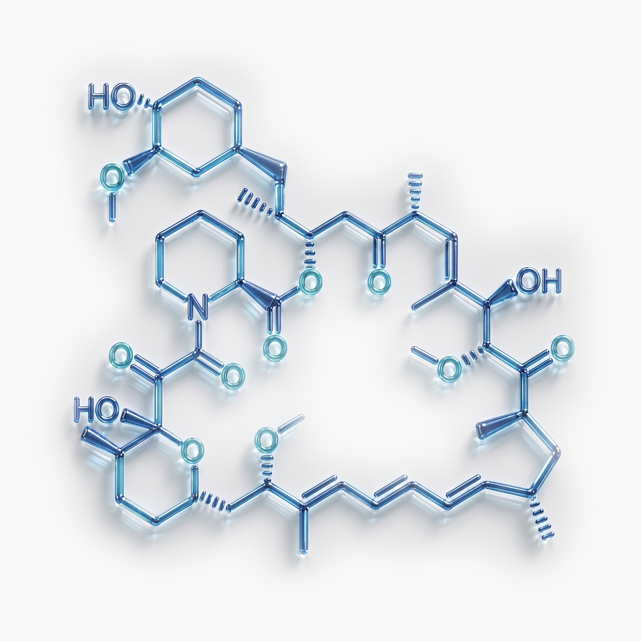
This biological understanding led directly to the approval of the first medicine shown to slow the progression of LAM. The drug sirolimus, previously approved to prevent the rejection of transplanted organs, was known to inhibit the mTOR pathway. Following clinical development, sirolimus was shown to suppress the growth and survival of abnormal LAM cells and slow the decline in lung function in many patients. Today, sirolimus remains the only FDA-approved therapy for LAM.
The MILES trial established sirolimus as a standard medical treatment for LAM and changed patient care worldwide. However, important unmet needs remained. Sirolimus does not eliminate the abnormal LAM cells, restore normal TSC1 or TSC2 function, or reverse established lung damage. Disease activity frequently resumes if treatment is discontinued, and some patients continue to experience disease progression despite treatment. Others are unable to tolerate long-term therapy because of treatment-related adverse effects.
These limitations demonstrate that important components of LAM biology remain untreated and support the need for complementary therapeutic approaches directed at additional disease pathways.
Beyond the mTOR pathway: Targeting VEGFR3
Increasing understanding of LAM biology has shown that abnormal activation of the mTOR pathway is only one component of the disease.
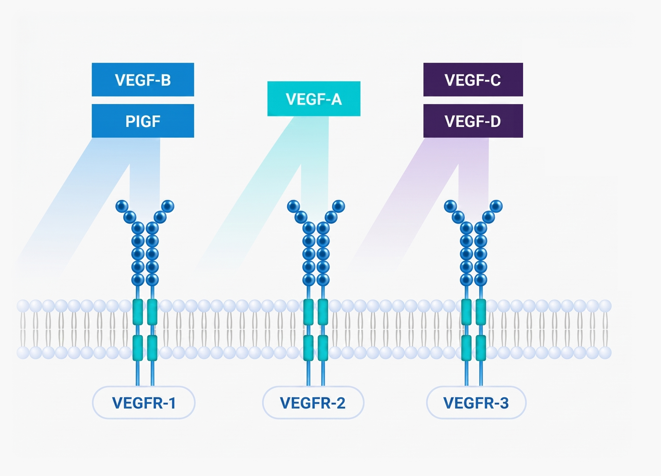
LAM cells do not simply proliferate. They also migrate through the lymphatic system, remodel lymphatic vessels, form lymphangioleiomyomas, contribute to chylous complications and progressively disrupt the normal architecture of the lungs. These processes are linked to a second major biological pathway involving vascular endothelial growth factors C and D (VEGF-C and VEGF-D) and their receptor VEGFR-3.
Under normal physiological conditions this pathway regulates the growth, maintenance and repair of the lymphatic system. In LAM, however, excessive activation of the VEGF-C/VEGF-D–VEGFR-3 pathway contributes to abnormal lymphatic remodelling, cellular migration and disease progression.
Unlike the mTOR pathway, which primarily regulates the growth and survival of abnormal LAM cells, the VEGF-C/VEGF-D–VEGFR-3 pathway regulates how those cells interact with the lymphatic system and surrounding tissues. Together, these complementary pathways drive much of the disease process observed in LAM.
This understanding has created a new therapeutic opportunity beyond mTOR inhibition.
Ceryvyn’s therapeutic strategy
Ceryvyn is developing a differentiated therapeutic approach based upon this biology. Its lead investigational therapy, CYV-101, is unique and has been designed to simultaneously trap both VEGF-C and VEGF-D, preventing activation of VEGFR-3 and interrupting one of the central biological pathways believed to contribute to disease progression. Unlike sirolimus, which suppresses abnormal LAM cell growth through inhibition of the mTOR pathway, CYV-101 is designed to target a complementary mechanism that regulates abnormal lymphatic biology.
By targeting a complementary biological driver of disease, this approach has the potential to complement existing mTOR inhibition and, if supported by future clinical studies, could provide the basis for a disease-modifying therapeutic strategy rather than one that primarily slows disease progression.
The company’s initial clinical development strategy is focused on inhaled delivery, enabling direct administration of therapy to the lungs while minimising systemic exposure. Because LAM is a systemic disease, demonstration of meaningful pulmonary benefit could provide the biological rationale for future evaluation of systemic administration to address extrapulmonary manifestations.
The next generation of clinical development
The understanding of LAM biology is changing not only how new therapies are designed, but also how they may be evaluated. Historically, clinical trials relied primarily on conventional pulmonary function tests such as FEV₁ and DLCO, the same endpoints used in the landmark MILES Trial. These measurements remain the international standard for assessing lung function and continue to play an important role in patient care.
However, conventional pulmonary function tests were originally developed for more common lung diseases such as asthma, chronic obstructive pulmonary disease (COPD) and idiopathic pulmonary fibrosis. They measure the overall consequences of lung damage rather than the biological processes responsible for it and provide only a single global assessment of lung function.
Ceryvyn’s development philosophy is to utilise the most informative and scientifically validated technologies available to understand both the biology of LAM and the effects of investigational therapies on that biology. As new technologies emerge and become scientifically validated, they have the potential to provide a more precise understanding of disease activity than has previously been possible. One of the most promising advances is hyperpolarized xenon-129 magnetic resonance imaging (Xe-129 MRI). Unlike conventional pulmonary function tests, advanced functional imaging such as hyperpolarized xenon-129 MRI has the potential to measure changes in regional lung biology rather than simply the downstream consequences of lung damage.
These technologies could enable earlier, more precise assessment of therapeutic response and support the next generation of clinical trial design in LAM. More sensitive assessments have the potential to support smaller and more efficient clinical trials, shorten the time required to evaluate promising therapies and provide a more detailed understanding of how investigational medicines influence the biological processes responsible for disease progression.
Ceryvyn believes that future therapeutic development should seek not only to target the biological drivers of LAM but also to measure those biological effects using the most advanced technologies supported by evolving scientific evidence and regulatory guidance.
Whether these advances ultimately become incorporated into future clinical trial designs or accepted regulatory endpoints will depend upon continued scientific validation and discussions with regulatory authorities. Nevertheless, the convergence of a deeper understanding of LAM biology, therapies directed at fundamental disease mechanisms and increasingly sophisticated methods of measuring biological response represents one of the most promising opportunities to accelerate the development of new treatments for LAM.
A growing global LAM network
Today, there are around 70 global LAM clinics informing patient databases, a biospecimen repository, and international conferences. These clinics are supported by family networks of people living with LAM.






Relevant publications
1.Lynn, E., et al. “Updated Prevalence of Lymphangioleiomyomatosis in Europe.” American Journal of Respiratory and Critical Care Medicine 209, no. 4. (2024): 456-459
2. Abbott, Gerald F., et al. “Lymphangioleiomyomatosis: Radiologic-Pathologic Correlation.” RadioGraphics 25, no. 3 (2005): 803–828.
3. Walkup, Laura L., et al. “Cyst Ventilation Heterogeneity and Alveolar Airspace Dilation as Early Disease Markers in Lymphangioleiomyomatosis.” Annals of the American Thoracic Society 16, no. 8 (2019): 1008–1016.
4. Bhattacharya, I, et al. “Assessment of Lung Structure and Regional Function Using 0.55 T MRI in Patients With Lymphangioleiomyomatosis.” Investigative Radiology 57, no. 3 (2022): 178–186.
5. Image from online presentation given by Nishant Gupta, MD, in partnership with The Lam Foundation. Retrieved from The LAM Foundation YouTube channel on 15/06/2026.
-
Get to know us
-
Talk to our team
-
Feel free to contact us!
Ceryvyn Therapeutics Limited
Ceryvyn Therapeutics Limited ABN 32 006 340 567.
An Australian biotechnology company (ASX: CYV) developing a biologic therapy for the rare disease LAM
(lymphangioleiomyomatosis).

Site Links
Contact Us
- Suite 1, Level 1, 117 Camberwell Road,
Hawthorn East VIC 3123
Subscribe to Updates
© 2026 Ceryvyn Therapeutics All rights reserved